
HONG KONG: First isolated in 1992 from Acanthamoeba polyphaga growing in a water tower in Bradford, UK, Mimivirus is one of the largest, most complex viruses known.
The Mimivirus’ structure is so similar to that of the amoeba that it was only identified as a virus in 2003.
The virus appears to be an icosahedral-shaped particle with a diameter of 400 nm and no envelope, surrounded by 80-nm-long fibrils.
It has a double-stranded DNA circular genome of about 800 kilobase pairs.
Both the genome and the virion size of the virus are larger than that of some small bacteria.
Researchers have been trying to determine the inner structure of this and other giant viruses to learn more about their origins.
For example, did they borrow genes over time from the host organisms they infect, like amoebas? Did they precede cell-based life or devolve from cell-based organisms?
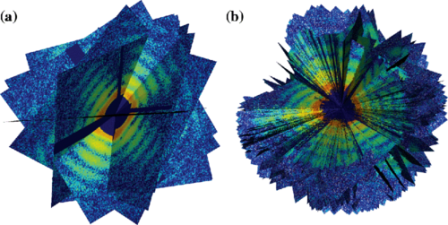
In the new study, scientists led by Dr Tomas Ekeberg of Uppsala University in Sweden sprayed a gas-propelled aerosol containing active Mimivirus virions in a thin stream into the X-ray laser beam, which scattered off the virions and produced light patterns on a detector that were recorded as diffraction images.
They then compiled hundreds of individual images from separate virions into a single 3D portrait showing the general shape and inner features of Mimivirus.
“We can see quite clearly that the inside of these viruses is not uniform,” said Dr Ekeberg, who is the lead author of the paper published in the journal Physical Review Letters.

Tesla driverless system to use updated radar technology
WASHINGTON: Electric carmaker Tesla announced Sunday it was upgrading its Autopilot software to use more advanced radar technology. In a...